|
 |
MYCOLOGICAL INSTITUTE
for the study of
|
 |
FUNGAL MOLD IN HUMAN HABITATIONS |
|
|
|
Instituut voor Studie van Schimmel in Menselijke Woningen |
|
Institut pour l'Etude de Moisissure Fongique dans Habitations Humaines |
|

|
|
Forschung Institut für Schimmelpilze in Innenräumen |
|
(NO CLAIM IS MADE TO ANY ORGINAL WORK) |
|
|
|
Aspergillosis is a non contagious disease of a number of animal species caused by fungi of the genus Aspergillus (usually A. fumigatus). Other potentially pathogenic species are A. flavus, A. nidulans, A. deflectus, A. niger and A. terreus . These fungi occur widely in nature and the usual mode of infection is by inhalation with the production of granulomatous lesions and nodules in the respiratory tract from which there may be dissemination to other tissues and organs.
Infections may also, but less frequently, begin in the alimentary tract. Disseminated aspergillosis originating from pulmonary or intestinal aspergillosis is a rare disease of dogs and cats. It is more common in immunosuppressed animals and in those under prolonged antibiotic treatment. Disseminated aspergillosis is probably most often diagnosed following necropsy. If diagnosed ante mortem it is treated long term with itraconazole or amphotericin B.
Nasal or paranasal aspergillosis is seen most commonly in the dog
Diagnosis - In general diagnosis is usually based on the isolation and identification of the Aspergillus species from appropriate clinical specimens and the demonstration of typical septate hyphae in biopsies or tissue sections. See treatment below.
|
|
|
|
|
Rottweiler crossbred dog with nasal aspergillosis
A Rottweiler crossbred dog with nasal aspergillosis due to Aspergillus fumigatus infection. Note the loss of pigment below the nostril on the worst affected side - this finding is suggestive of a diagnosis of chronic nasal aspergillosis in the dog. |
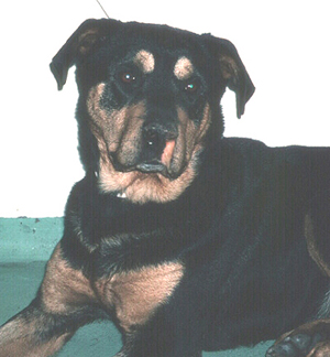
(© Dr. R. Mallik, Sydney, Australia)
|
|
Aspergillus sinusitis in a dog
Long nosed dogs are at relatively high risk of Aspergillus sinusitis as shown in this example
|
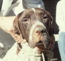
(© Dr. R. Mallik, Sydney, Australia)
|
|
|
|
Canine Nasal Aspergillosis
Labrador retriever with nasal infection by Aspergillus terrus. With early diagnosis and intervention this state could have been avoided. |
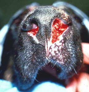
(© Dr. R. Mallik, Sydney, Australia |
|
|
English Pointer with nasal aspergillosis treated by topical enilconazole injected through surgically inserted indwelling plastic tubes. (see treatment)
|
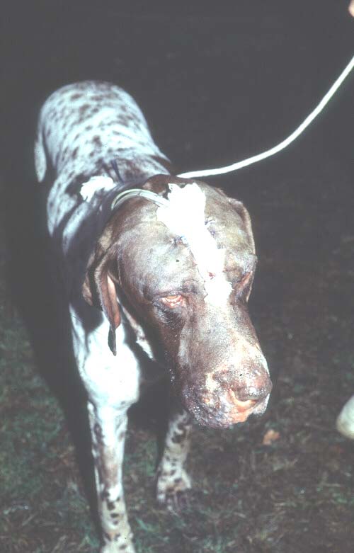
(© Dr. R. Mallik, Sydney, Australia)
|
|
|
HEART INFECTION
|
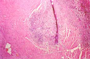
Myocardial aspergillosis When the heart is involved in an inflammatory process, often caused by Aspergillus sp. G ranuloma within the myocardium(middle layer of heart muscle) of dog. Commonly resulting in "enlarged heart" leading to death.
|
(© Dr. Michael Day, University of Bristol)
|
|
EYE INFECTION
|
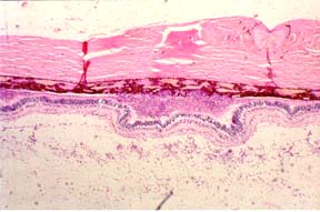
Retinal Aspergillosis (dog J) Section of retina from a German shepherd dog with disseminated aspergillosis. Fungal hyphae and inflammatory cells are found within the vitreous(eye).
|
(© Dr. Michael Day, University of Bristol)
|
|
|
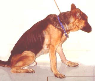
WASTING
A 2 year old, male German shepherd dog with disseminated aspergillosis due to Aspergillius terreus. The marked loss of condition of this dog occurred within two months of initial diagnosis
(© Dr. Michael Day, University of Bristol)
|
|
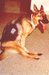
PARAPALEGIA
A 2 year old, female German shepherd dog with disseminated aspergillosis due to Aspergillius terreus. There is muscle wasting and paraplegia due to discospondylitis involving T13-L1.
(© Dr. Michael Day, University of Bristol)
|
|
|
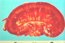
KIDNEY INFECTION
| Saggital section of kidney from a German shepherd dog with disseminated aspergillosis. There are granulomata within the medulla, and fungal material within the renal pelvis. Renal involvement in canine disseminated aspergillosis is common, and the demonstration of fungal hyphae within urine sediment is a useful screening test. |
(© Dr. Michael Day, University of Bristol)
|
|
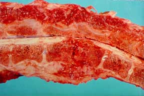
DISCOSPODYLITIS-SPINAL CORD
|
Discospondylitis Saggital section of the vertebral column of a dog with discospondylitis as part of disseminated aspergillosis due to Aspergillus terreus.
|
(© Dr. Michael Day, University of Bristol)
|
|
|
PULMONARY DISEASE
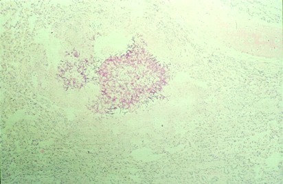
Pulmonary aspergillosis with periodic acid Sciff ( PAS) staining section of lung from a feline (cat).
|
(© Dr. Michael Day, University of Bristol |
|
|
MENINGITIS
Meningitis (dog R)Grocott-stained serial section of the previous slide for dog R reveals the presence of fungal material at the centre of each microgranuloma. These lesions were not cultured, but Aspergillus spp . was identified by immunohistochemical examination using a panel of specific antisera. There was no evidence of systemic involvement in this dog. This case was reported in Veterinary Record 1995, 136: 38-41.
|

(© Dr. Michael Day, University of Bristol |
|
|
|
Books by Dr Michael Day BSc, BVMS(Hons), PhD, FASM, Dipl. ECVP, MRCPath, FRCVS
Dr Day is Reader in Veterinary Pathology at the University of Bristol. He is a diagnostic histopathologist and runs a clinical immunology diagnostic service. His research interests cover experimental models of autoimmunity and a range of companion animal immune-mediated and infectious diseases. |
|
|
ORDER
|

By Michael J. Day An excellent resource for veterinarians in practice and in training, Clinical Immunology of the Dog and Cat provides information on such topics as immunodeficiency disease, immune system neoplasia, and immunotherapy.
Illustrations, including 670 color diagrams and photographs form clinical cases, cover radiography, endoscopy, cytology, gross and microscopic pathology, immunopathology, and electron microscopy.
A list of abbreviations and references for additional reading further complement this reference book. Also, a set of symbols, defined in a key, is used to consistently represent immunological molecules and cells in diagrams throughout the book.
|
|
ORDER
|
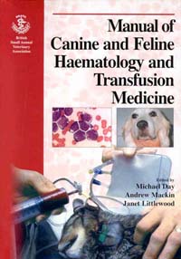
By Michael J. Day, Andrew Mackin, Janet D. Littlewood
Bridges the disciplines of clinical pathology, internal medicine, and critical care in a single volume. The manual is divided into the three following sections including haematology, haemostasis, and transfusion medicine.
About the Author: Michael J. Day, BSc, BVMS (Hons), PhD, FASM, Dipl ECVP, MRDCPath, FRCVS, is senior lecturer in veterinary pathology, Department of Pathology and Microbiology, School of Veterinary Science, University of Bristol, UK.
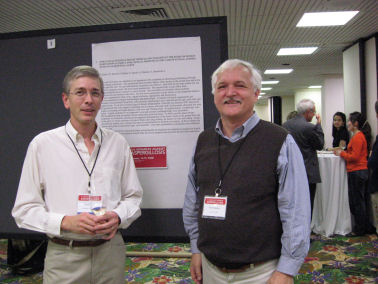
Day and Dumanov Aspergillosis 3rd Miami, Jan 15-20,2008 |
|

Page dedicated to "CHAT" beloved cat of the Mycological Research Insititute. 2000?- Jan. 3, 2008
|
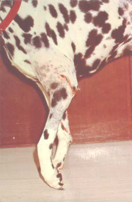
OSTEOMYELITIS
Draining sinus tract on left forelimb of a 4 year old, female Dalmatian with disseminated aspergillosis. There was an underlyi
ng osteomyelitis of distal humerus from which Aspergillus terreus was cultured. |
|
|
LYMPH GRANULOMA
Lymph node granuloma. Section of lymph node granuloma from a German shepherd dog with disseminated Aspergillosis stained for canine IgA by immunofluorescence. The fungal hyphae within the centre of the lesion have surface IgA, and IgA-bearing plasma cells are present within the surrounding inflammatory infiltrate. This apparently 'inappropriate' usage of IgA in a systemic immune response, may be part of a wider problem of 'IgA dysregulation' in German shepherd dogs, and may be one factor predisposing dogs of this breed to disseminated aspergillosis. |
|
|
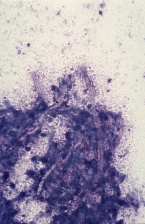
Domestic crossbred cat with disseminated aspergillosis
 (© Dr. R. Mallik, Sydney, Australia (© Dr. R. Mallik, Sydney, Australia
Diff Quik stained squash preparation of material obtained from thoracotomy of a 3 year old domestic crossbred cat with invasive Aspergillus fumigatus infection. The cat had marked enlargement of the hilar lymph nodes that resulted in a partial tracheal obstruction. This smear was made from portions of the hilar lymph node resected at thoracotomy. Magnification x 132.
|
|
Disseminated aspergillosis in a German Shepherd 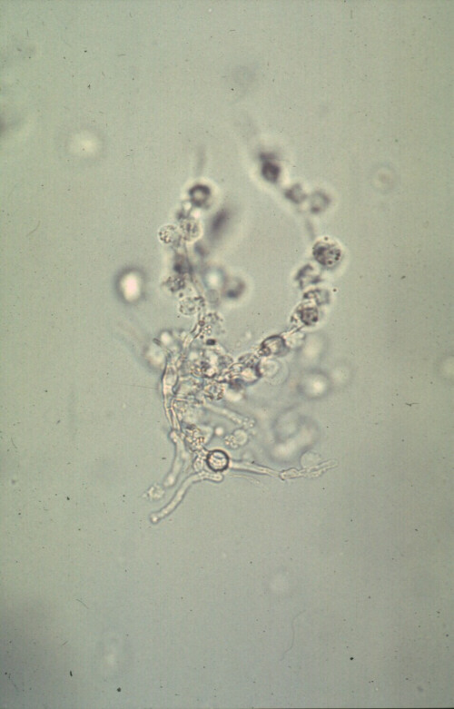 (© Dr. R. Mallik, Sydney, Australia (© Dr. R. Mallik, Sydney, Australia
Hyphae of Aspergillus terreus in the urine of a young German Shepherd dog with disseminated aspergillosis. White blood cells are present in addition to the fungal hyphae. Wet preparation of urine; magnification x 132.
|
|
|





 (© Dr. R. Mallik, Sydney, Australia
 (© Dr. R. Mallik, Sydney, Australia